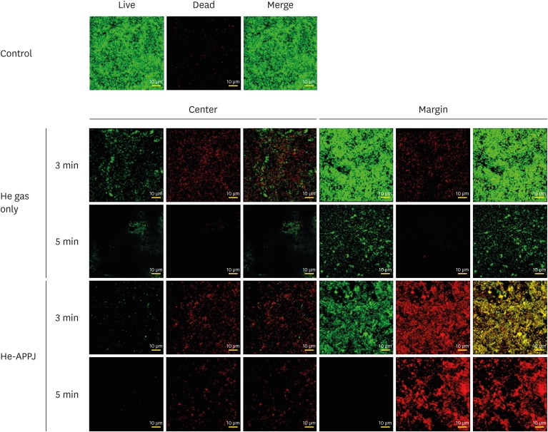
Figure 3. Confocal laser scanning microscopy images of the central and marginal areas after He-APPJ application. Heavy live bacterial accumulation on the discs was observed in the control group. In the He gas–only treatment group, the amount of bacteria was higher in the central area than in the marginal area in the 3-minute treatment group. In the 5-minute treatment group, the amount of bacteria decreased in both the central and marginal areas, although more live bacteria were observed in the marginal area. in the He-APPJ treatment group, the amount of bacteria was lower in the central area than in the marginal area in both the 3-minute and 5-minute treatment groups. Only a few viable bacteria were observed in the central area of the 3-minute treatment group. In the marginal area, a mixture of live and dead bacteria was found, whereas most bacteria were dead in the 5-minute treatment group.
He: helium, He-APPJ: helium atmospheric-pressure plasma jet.

