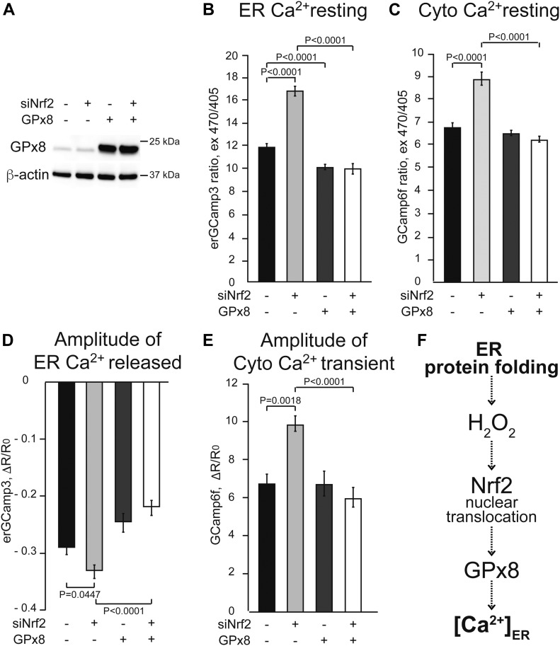
Figure 4. Expression of GPx8 normalizes ER calcium dysregulation, downstream of Nrf2 signaling.
HeLa cells were transfected with GPx8 (+) or pcDNA3 (−) for 48 h, and then with Nrf2 siRNA (+) or control siRNA (−) for 24 h. All data are presented as mean ± SEM. (A) Representative Western blot of GPx8 overexpression and β-actin as loading control. (B) Resting ER calcium measured by erGCaMP3. n = 317, 186, 253, 337 cells from three sets of experiments. P values between indicated groups by Kruskal–Wallis test with Dunn’s correction are shown. (C) Resting cytosolic calcium levels measured with GCaMP6f. n = 325, 331, 505, 402 cells from three sets of experiments. P values between indicated groups by Kruskal–Wallis test with Dunn’s correction are shown. (D) ER calcium released upon 100 μM histamine stimulation, measured by drop in erGCaMP3 fluorescence. n = 36, 36, 52, 77 cells from three sets of experiments. P values between indicated groups by paired one-way ANOVA test with Sidak’s correction are shown. (E) Cytosolic calcium transient peak induced with 100 μM histamine, measured by GCaMP6f. n = 41, 49, 61, 73 cells from three sets of experiments. P values between indicated groups by Kruskal–Wallis test with Dunn’s correction are shown. (F) Proposed model linking ER protein folding and ER calcium through H2O2 signaling, Nrf2 nuclear translocation, and GPx8 expression.

