Fig. 1.

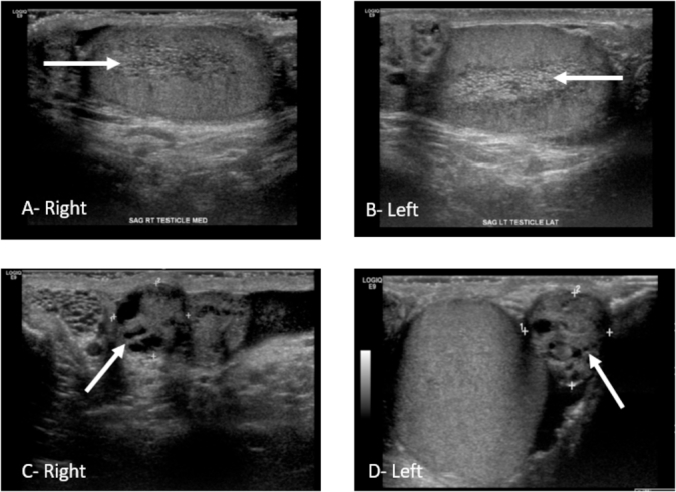
Ultrasound Images. Right and Left Testicles noted dilated rete testes (Arrows-Images A, B); Bilateral epididymal lesions with mostly solid components and small cystic areas consistent with cystadenomas (Arrows-Images C, D).

Ultrasound Images. Right and Left Testicles noted dilated rete testes (Arrows-Images A, B); Bilateral epididymal lesions with mostly solid components and small cystic areas consistent with cystadenomas (Arrows-Images C, D).