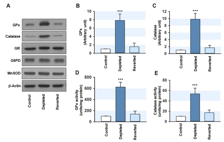
Fig. 3. Effect of mitochondrial DNA (mtDNA) depletion on the expression and activity of glutathione peroxidase (GPx) and catalase.
The total cell lysates were prepared in the control, mtDNA-depleted (Depleted) and -reverted (Reverted) myoblasts. (A–C) The expression levels of GPx and catalase were analyzed by immunoblotting. The densities were normalized to the β-actin signals, and the relative intensities are expressed in arbitrary units, where the intensity of the control was set to one. (D) The total GPx activity was measured using the coupled enzyme procedure with glutathione reductase. The specific activity was calculated using the extinction coefficient obtained from the NADPH standard. (E) The total catalase activity was measured by monitoring the decomposition of 10 mM H2O2 at 240 nm in a medium. One unit of catalase decomposes 1 mM of H2O2 per min. The values are expressed as the mean ± SEM from four independent experiments. GR, glutathione reductase; SOD, superoxide dismutase. ***p < 0.001 vs. control.

