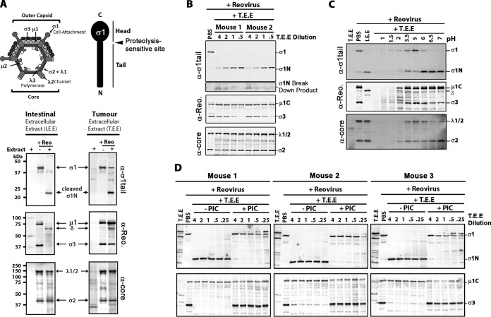
FIG 1.
Breast tumor extracellular protease activity cleaves reovirus σ1 protein but does not produce ISVPs. Extracellular contents of murine breast tumors (tumor extracellular extract, or T.E.E.) were used to mimic the tumor microenvironment. To mimic the intestinal environment, the protease-rich luminal contents were obtained from murine intestines (intestinal extracellular extract, or I.E.E.). (A, top) Diagrammatical depiction of reovirus outer capsid and core proteins, along with a diagram of σ1 tail and head domains. (Bottom) Reovirus (Reo) was treated with T.E.E. or I.E.E. at 37°C for 24 h, and viral proteins were subjected to Western blot analysis using antibodies against σ1-tail, outer capsid proteins (polyclonal antireovirus serum), or viral core proteins (polyclonal antireovirus core serum). (B) As described for panel A, but the activity of T.E.E. from two additional and independent murine breast tumors was assessed. (C) Reovirus was treated with T.E.E. under different pH conditions, ranging from pH 1 to pH 7. (D) Reovirus was treated with T.E.E. from 3 murine breast tumors in the absence or presence of protease inhibitor cocktail (PIC).

