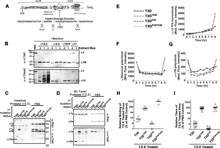
FIG 6.
Mutation in the σ1 neck domain can overcome σ1 proteolysis by breast cancer metalloprotease. (A) Diagrammatic depiction of σ1 with the protease-hypersensitive neck domain. (B) Reovirus was treated with either T.E.E., I.E.E., chymotrypsin (CT), or trypsin (TRYP) for 24 h at 37°C and subjected to Western blot analysis with tail (top)- or head (bottom)-specific antibodies. (C and D) CsCl-purified T3D, T3DT249I, T3DS18I, or T3DS18I/T249I was treated with PBS or with I.E.E. (C) or T.E.E. (D) for 24 h at 37°C. Western blot analysis with both polyclonal antireovirus antibodies and σ1N-specific antibodies demonstrates the levels of full-length σ1 and σ1N. (E to G) Reovirus infection dynamics of T3D, T3DT249I, T3DS18I, and T3DS18I/T249I viruses. Flow cytometry was used to evaluate expression of reovirus proteins λ2 (E), σ3 (F), and σ1N (G), from 0 to 8 h postinfection. (H) Binding assay as described for Fig. 3 with reovirus mutant T3D, T3DT249I, T3DS18I, or T3DS18I/T249I treated with T.E.E. on L929 cells. (I) Plaque titration of T.E.E.-treated reovirus mutants (T3D, T3DT249I, T3DS18I, or T3DS18I/T249I) as described for Fig. 2.

