FIG 2.
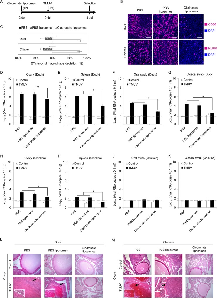
Roles of monocytes/macrophages in TMUV infection in vivo. (A) Scheme depicting the design of the experiment. Monocytes/macrophages were depleted by intraperitoneal injection of clodronate liposomes. (B) Representative pictures of immunofluorescence staining for monocyte/macrophage markers. The efficiency of monocyte/macrophage depletion by clodronate liposome injection was examined in sections of spleen samples 2 days after clodronate liposome injection using rabbit anti-CD68 polyclonal antibody for duck samples and mouse anti-chicken monocyte/macrophage-PE clone KUL01 for chickens. Equal volumes of a control liposome suspension and PBS were used as controls. Scale bars, 30 μm. (C) The numbers of monocytes/macrophages were counted, and the reduction rates were calculated. Data are presented as the mean ± SEM (n = 3). (D to M) At 3 days post-intravenous injection of 500 μl of virus specimens at 1 × 10⁵ PFU per ml in ducks and chickens preinjected with clodronate liposomes, control liposome suspension (PBS liposomes), or PBS 2 days prior to infection, ovaries were harvested, and oral swabs and cloaca swabs were collected. The level of viral RNA was detected by RT-qPCR. Samples collected from uninfected animals are indicated as “control.” A dashed line indicates the limit of detection. (L and M) Representative H&E staining of ovary section slides at a higher magnification with arrows depicting areas of damage are shown. The data in panels D to K are presented as the mean ± SEM. Asterisks indicate significant differences (n = 3, P < 0.05). Scale bars in panels L and M, 200 μm.

