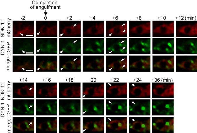
Figure 2.
Both NDK-1 and DYN-1 are enriched on the surface of extending pseudopods and maturing phagosomes and partially overlap on the phagosomal surface. Time-lapse recording of DYN-1::GFP and NDK-1::mCherry, which are coexpressed in engulfing cells during the engulfment and degradation of cell corpse C3 in a wild-type embryo. “0 min” indicates the moment a nascent phagosome is just formed. Arrows indicate a few regions on extending pseudopods or the phagosome in which colocalization of GFP and mCherry is observed. Ten phagosomes were monitored by time-lapse recording, and the partial colocalization was observed from all of these phagosomes. Scale bar, 2.5 µm.

