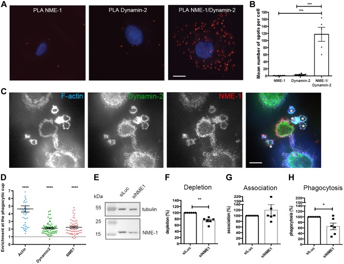
Figure 4.
Role of NME1 in phagosome formation in human. Primary human macrophages were differentiated from blood monocytes. A, B) PLA was performed with anti-NME1 and anti-Dynamin antibodies. Images were acquired (A), and quantification of spots was performed and analyzed by ANOVA (B). Phagocytosis of zymosan was performed for 10 min before fixation, permeabilization, and labeling with phalloidin Alexa 635 and anti-NME1 followed by Cy3-coupled anti-mouse IgG antibodies and anti-Dynamin antibodies followed by Alexa 488–coupled anti-rabbit IgG antibodies. One confocal section is shown. C) Asterisks label the phagocytic cups. D) Protein enrichment in phagocytic cups was determined as described in Materials and Methods. E–H) Cells were treated with siRNA against NME1 (siNME1) or luciferase (siLuc) as a control for 72 h before cell lysis and Western blot analysis (E, F) or phagocytosis assay with IgG-opsonized red blood cells (G–H). Association (G) and phagocytosis (H) efficiencies were calculated as indicated in Materials and Methods and expressed as a percentage of control cells treated with siLuc for 6 different donors. Error bars represent sem. For depletion, association, and phagocytosis, we used 1-sided Student’s t tests. Scale bar, 5 µm. Error bars represent sem. *P < 0.05, **P < 0.01, ***P < 0.001, ****P < 0.0001.

