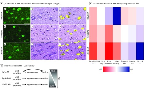
Figure 1. Selective Vulnerability of the Nucleus Basalis of Meynert (nbM) and Corticolimbic Structures to Neurofibrillary Tangles (NFTs) Among Neuropathologic Subtypes of Alzheimer Disease (AD).
A. Thioflavin S microscopy (a, c, and e) shows greater NFT accumulation in the nbM of hippocampal sparing (HpSp) AD (a) compared with typical AD (c) and limbic predominant (limbic) AD (e). Hematoxylin-eosin–stained sections of the nbM (b, d, and f) were digitally quantified (b′, d′, and f′, respectively). Fewer neurons are observed in HpSp AD (b) compared with typical AD (d) and limbic predominant AD (f). Scale bar represents 50 μm. B. Heatmap of differences calculated between brain region of interest and the nbM, as exampled by the more severe involvement of the entorhinal cortex compared with the nbM in limbic predominant AD, shown in warmer colors, and the less severe involvement of the hippocampus (Hipp) in HpSp AD compared with the nbM, shown in cooler colors. C. We hypothesize that, although both the nbM and entorhinal cortex (ctx) are involved early among AD subtypes and across aging, the cortex may be more vulnerable in HpSp AD. By contrast, the pattern of greater vulnerability of limbic structures is manifested in both limbic predominant AD and perhaps as a function of older age. LOAD indicates late-onset AD; YOAD, young-onset AD.

