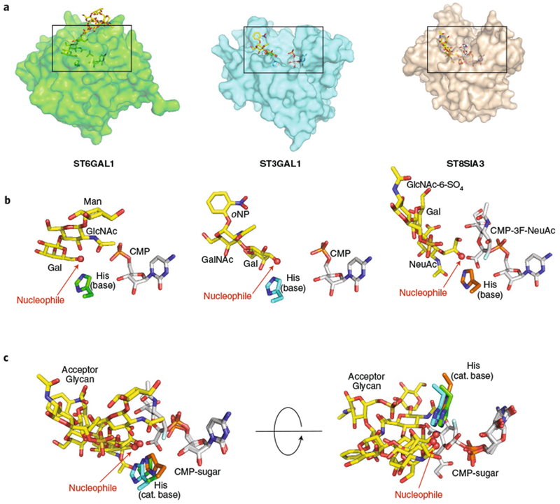
Fig. 4 |. Structural gallery of eukaryotic GT29 sialyltransferases as acceptor-bound complexes.

a, Three eukaryotic GT29 sialyltransferase structures (GT-A fold (variant 2)) were aligned using Coot88 (transparent surface) with bound donor analogs (white sticks), acceptor analogs (yellow sticks), and catalytic bases (sticks). Structures include ST6GAL1 (PDB: 4JS235), ST3GAL1 (PDB: 2WNB37), and ST8SIA3 (PDB: 5BO938). Boxes represent the regions of the respective structures shown in b. b, Enlarged views of the bound ligands are displayed in the same orientation and coloring as in a, with individual donors, acceptors, catalytic bases labeled as in Fig. 3. For ST6GAL1, the large Gal2GlcNAc2Man3GlcNAc2-Asn ligand has been simplified to the terminal Gal-β-1,4-GlcNAc-β-1,2-Man acceptor. c, Overlay of the three bound ligand complexes in two different orientations.
