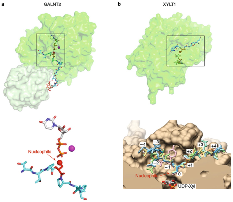
Fig. 6 |. Glycosyltransferases recognizing linear peptides.

a, GALNT2 (PDB: 5AJP69). Enzyme represented in transparent surface representation, with the catalytic domain in green and lectin domain in light green. Donor nucleotide, white sticks; acceptor peptide, cyan sticks; GalNAc on acceptor peptide, red sticks; manganese, magenta sphere. Lower panel: enlarged view of the bound ligands are displayed in the same orientation and coloring as that shown on top with individual molecule components labeled. Acceptor amino acid is shown in red sticks, with the acceptor oxygen as a red sphere. b, XYLT1 (overlay of PDB IDs 6EJ7 and 6EJ839). The catalytic domain of the enzyme is represented in transparent surface representation in green. Donor nucleotide, white sticks; acceptor peptide, cyan sticks. Lower panel: enlarged cut-through view of the active site in tan with donor UDP-Xyl at the bottom, showing superposition of eight acceptor peptides in sticks (reproduced with permission from ref. 39). Boxes in the upper panels represent the regions of the respective structures shown in the lower panels.
