
Fig. 7 |. Domain specific GTs (POFUT1, POFUT2, and POGLUT1) with substrates.
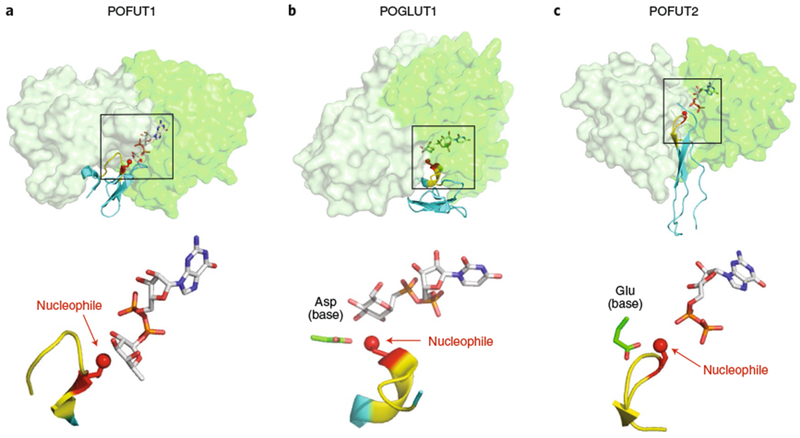
a, POFUT1 (overlay of PDB IDs 5KXH and 5KY320). b, POGLUT1 (overlay of PDB IDs 5L0R and 5L0U43). c, POFUT2 (PDB: 5FOE42). Upper panels: all three GT-B fold enzymes are displayed in transparent surface representations with A domains in light green and B domains in darker green. Folded domains (EGF repeats for POFUT1 and POGLUT2; TSR for POFUT2) are in cyan cartoon with respective consensus sequences in yellow. Acceptor amino acid is shown in red sticks, with the acceptor oxygen as a red sphere. Donor nucleotides, white sticks. Note that the POGLUT1 structure has the donor analog UDP-CH2-Glc. Lower panels: enlarged views of the bound ligands are displayed in the same orientation and coloring as the upper panels, with individual molecule components labeled. Boxes in the upper panels represent the regions of the respective structures shown in the lower panels.