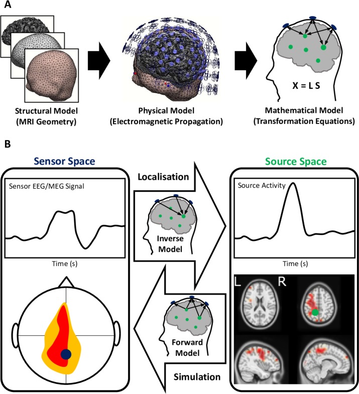
Figure 2.
Electromagnetic source imaging using electroencephalography and magnetoencephalography (EEG/MEG) source localisation (A) building models of source and sensor activity and (B) forward versus inverse transformation of signals between the sensors and brain sources. (A) MRI geometry is used for developing structural models of the brain, corticospinal fluid and scalp (among many layers) that are in-between the brain sources generating the neuroelectric activity and electrodes/sensors. The structural model when used together with physical electromagnetic properties of the tissue materials and the governing equations of electromagnetic propagation forms a physical model. The physical model is solved and formulated for the discrete finite number of the modelled sources of activity in the brain (usually about thousands), as well as the EEG/MEG sensors used during the data acquisition (usually a few hundreds). The mathematical model X=LS is a multivariate relationship between the sensor activity (X), source activity (S) and the mathematical model (L). (B) This mathematical model can forward-transform the simulated source activities to the sensors, as well as project the recorded sensor activity to localise the underlying brain sources using the constructed inverse model.

