Figure 2.
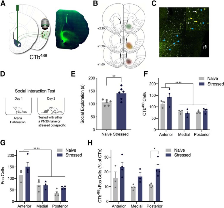
Exposure to stressed PN30 conspecifics elicited greater Fos immunoreactivity in IC → NAc neurons than exposure to naive PN30 conspecifics. A, Schematic depicting unilateral 300 nl injections of the retrograde tracer CTb488 in the right hemisphere NAc (left) and representative image of CTb488 in NAc (right: green = CTb488, blue = DAPI). B, CTb488 expression in NAc: each experimental rat is represented in a different color. C, CTb488- and Fos-positive neurons in IC. Image is psuedocolored; white indicates Fos, yellow is CTb488, blue is colabeled cells. rf, Rhinal fissure. D, Diagram of behavioral procedures. Six experimental rats explored naive PN30 conspecifics and six explored stressed PN30 conspecifics. E, Mean (with individual values) social exploration time. Experimental rats presented stressed PN30 conspecifics engaged in more social exploration than rats presented naive PN30 conspecifics (p = 0.008). F, Counts of CTb488-positive cells in anterior, medial, and posterior IC. More CTb488-positive cells were observed in anterior IC than medial or posterior IC (p < 0.0001); there was no effect of conspecific stress on the number of CTb488-positive cells at any ROI. G, Counts of Fos-positive cells in each IC region. More Fos-positive cells were observed in anterior IC than medial or posterior (p < 0.0001); there was no effect of conspecific stress on the number of Fos-positive cells at any ROI. H, Counts of colabeled CTb488 and Fos-positive neurons. A main effect of stress was observed (p = 0.0008), and experimental rats that explored stressed PN30 conspecifics expressed more Fos immunoreactivity in CTb488-positive neurons in posterior IC (p = 0.015) compared with experimental rats who explored naive PN30 conspecifics. *p < 0.05, **p < 0.01, ****p < 0.0001.

