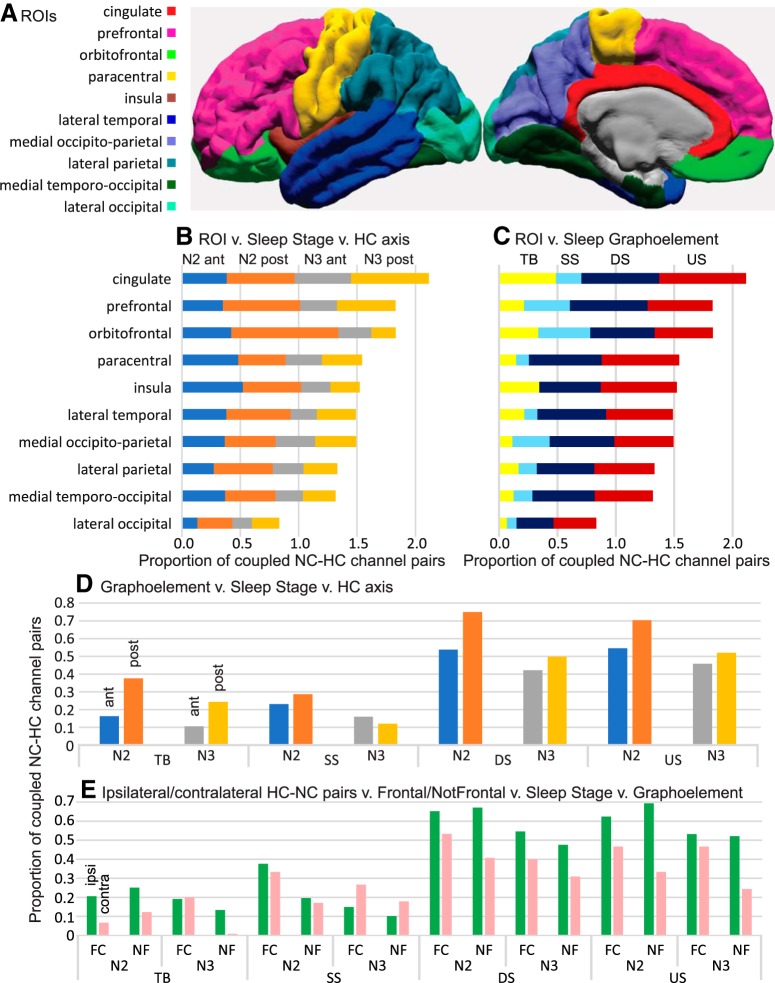
Figure 5.
Strength of HC-NC association varies across NC regions (B,C,E), NREM stages (B,D,E), GE types (C–E), and anterior versus posterior (D) or ipsilateral versus contralateral (E) SWR sources. A, Map of NC ROIs. B, C, Mean proportions of significant HC-NC channel pairs in different NC regions. Channels with significant (as evaluated in Fig. 2) HC-NC co-occurrences are more common in frontocentral (FC) than nonfrontal (NF) cortex (B–D), N2 than N3 (B,D,E), DS and US than TB and SS (C–E), with posterior than anterior HC-SWRs (D), and with ipsilateral than contralateral HC-SWRs (E). Within these general patterns, interactions can be observed, such as the relatively high co-occurrence of orbitofrontal GE with pHC-SWRs in N2 (B). Proportions are relative to total channels in each ROI (B,C), all ROIs (D), or frontocentral or nonfrontal ROIs in the ipsilateral versus contralateral hemisphere (E). For correspondence of ROIs to FreeSurfer labels, and their amalgamation to frontocentral and nonfrontal, see Extended Data Table 3-1.

