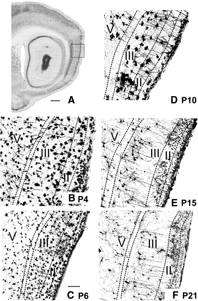
Figure 3.

Postnatal development of morphological properties of neurons in MEC. The postsynaptic side. Golgi-stained material showing the neuronal morphology at different postnatal days. A, Sagittal section of a P20 animal stained with NeuN as a marker for neuronal somata. Square represents the approximate position of the Golgi images for B–F. Scale bar, 1.5 mm. High magnification of a piece of MEC stained with Golgi (for details, see Materials and Methods) of a P4 (B), P6 (C), P10 (D), P15 (E), and P21 (F) animal. The neurons appear immature from P4 to P10 and adult-like starting at P15. Apical dendrites can be seen radiating from the deep layers to the pial surface in the 2 older animals. Neurons in superficial layers of the youngest animals (B) appear substantially larger than in older animals; this is due to an incubation artifact resulting in too dense silver deposits. Both neuronal stains in younger tissue (Nissl) as well in the intracellular fill data corroborate that the neurons at these young postnatal ages are not larger. Scale bar: (in C), B–F, 200 μm.
