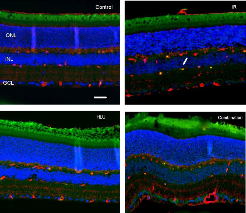
Figure 2:
Apoptosis based on terminal deoxynucleotidyltransferase dUTP nick-end labeling (TUNEL) staining of retinal tissue. i) Control, ii) Proton irradiation (IR) at 50cGy, iii) Hindlimb unloading (HLU), iv) Combination. TUNEL-positive cells were identified with green fluorescence, endothelium was stained with lectin (red). The nuclei of photoreceptors were counterstained with DAPI (blue). TUNEL-positive cells that were located within red lectin-labeled endothelium were identified as TUNEL-positive endothelial cells (ECs). ONL: outer nuclear layer, INL: inner nuclear layer, GCL: ganglion cell layer. Arrow: TUNEL-positive EC. In the control retinal tissue, only sparse TUNEL-positive cells were found. In the retina from mice exposed to 50 cGy of proton and HLU, TUNEL-positive labeling was apparent in the endothelial cells. Scale bar =50 μm. Green auto-fluorescent was noted in the outer layers.

