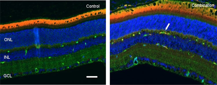
Figure 4:
Representative micrographs of retina sections evaluated for endothelial nitric oxide synthase (eNOS) expression at 30 days after proton radiation and/or hindlimb unloading (HLU). i) Control, ii) Combination. Endothelial NO synthase (eNOS) positive cells were identified with red fluorescence, endothelium was stained with lectin (green). The nuclei of photoreceptors were counterstained with DAPI (blue). eNOS-positive cells that were located within red lectin-labeled endothelium were identified as eNOS-positive endothelial cells (ECs). ONL: outer nuclear layer, INL: inner nuclear layer, GCL: ganglion cell layer. Arrow: eNOS-positive EC. In the non-irradiated retinal tissue, only sparse eNOS-positive cells were found. In the retina from mice exposed to 50 cGy of proton radiation combined with HLU, eNOS-positive labeling was apparent in the ECs. Scale bar =50 μm. Red auto-fluorescent was noted in the outer layers.

