Figure 7.
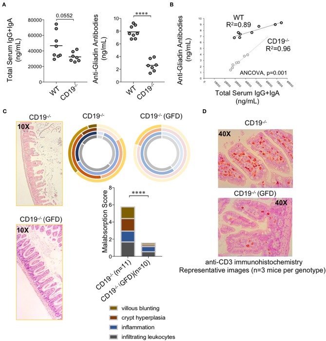
Malabsorption in CD19−/− mice is gluten-sensitive. (A) Total serum IgG and IgA and total AGA antibody titers are shown. (B) Correlation between AGA antibody titers and total IgG/IgA titers in WT and CD19−/− mice. (C) Malabsorption scoring for GFD-treated CD19−/− mice compared to animals reared on a gluten-containing mouse chow. (D) Representative anti-CD3 IHC staining of ileal T cells in CD19−/− mice treated with GFD vs. controls. (A) Student's t-test, **** = p < 0.0001. (C) Result of Two-way ANOVA (row factor = genotype), **** = p < 0.0001.

