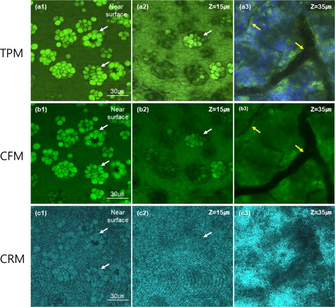
Figure 2.
3D TPM, CFM and CRM images of an ex-vivo mouse conjunctiva tissue. (a–c) moxifloxacin-based TPM and CFM images, and an CRM image of the same mouse conjunctiva tissue respectively. Three en-face images at different depths of the near surface, 15 μm and 35 μm deep from the surface are presented. The first two superficial images showed the epithelium and the last one image showed the substantia propria. Green color in both TPM and CFM images represented moxifloxacin fluorescence and blue color in the TPM image represented SHG. White and yellow arrows marked goblet cell clusters and blood vessels, respectively.

