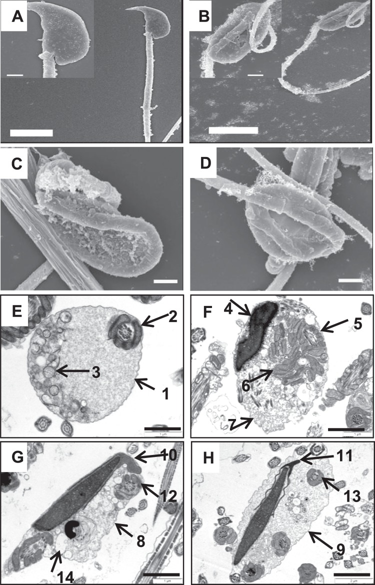
Figure 2.
Ultrastructure analyses of WT and Ncoa5+/− sperm. (A–D) Representative SEM analytic images of WT (A) and Ncoa5+/− epididymal sperm (B–D). Inserts are the higher magnification images of the head and neck region of the sperm. Scale bars: 5 μm (A,B); 1 μm (C,D); inserts: 1 μm. (E–H) Representative TEM analytic images of WT (E) and Ncoa5+/− epididymal sperm (F–H). Scale bars: 1μm. Cross-section of the cytoplasmic droplet (CD) of a WT spermatozoon (E). 1: Outer membrane of the CD; 2: middle piece of the sperm containing mitochondrial sheath and axoneme; 3: vacuoles within the CD; Cross-sections of Ncoa5+/− bent sperm head and neck region wrapped around by the tail (F–H). 4: nucleus; 5: middle piece containing mitochondria and axoneme wrapped around the bent head and neck; 6: disrupted mitochondria of the middle piece; 7, 8 and 9: outer membrane of the droplet; 10 and 11: acrosome; 12 and 13: wrapped-around middle piece containing mitochondria and axoneme; 14: large membranous vacuoles.

