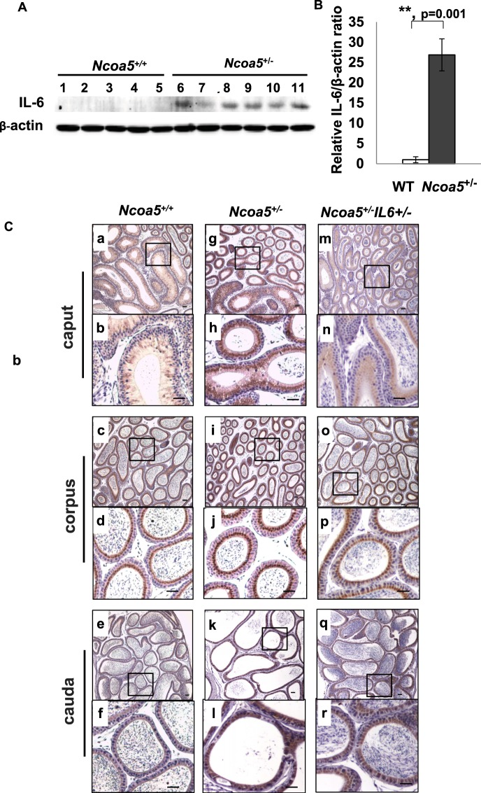
Figure 4.
IL-6 expression in epididymis of WT and Ncoa5+/− male mice. (A) Western blot analysis of IL-6 expression in epididymis of age-matched 13–15 months old WT (n = 5, lanes 1–5) and Ncoa5+/− (n = 6, lanes 6–11) male mice using anti-IL-6 and anti-β-actin antibodies. Each lane represents a single tissue lysate of the whole epididymis from a mouse. Cropped blots from the same gel are shown. Uncropped blots are presented in Supplementary Fig. S4. The same blot membrane was stripped and blotted with the same method except without adding the primary antibodies as control (Supplementary Fig. S4C). (B) Quantification of data from (A). n = the number of mice. **P < 0.01. Error bar ± SEM. (C) Representative IL-6 IHC staining of WT, Ncoa5+/− and Ncoa5+/−Il-6+/− male mouse epididymides. Immunohistochemical analysis was performed on epididymis from 4.5 months old WT (n = 2), Ncoa5+/− (n = 3) and Ncoa5+/−Il-6+/− (n = 3) male mice using an anti-IL-6 antibody. n = the number of mice. Representative IL-6 IHC staining images of WT (a–f), Ncoa5+/− (g–l) and Ncoa5+/−Il-6+/− (m–r) epididymal caput (segments 1–2) at lower (a, g and m) and higher (b, h and n) magnification, corpus (segments 7) at lower (c, i and o) and higher (d, j and p) magnification, and cauda (segments 8–9) at lower (e, k and q) and higher (f, l and r) magnification. Black squares specify the areas that are magnified and displayed as high magnification pictures. Scale bars: 50 µm. Immunohistochemical analysis without using IL-6 antibody was performed on epididymis from an Ncoa5+/− mouse and a representative image is shown in Supplementary Fig. S5.

