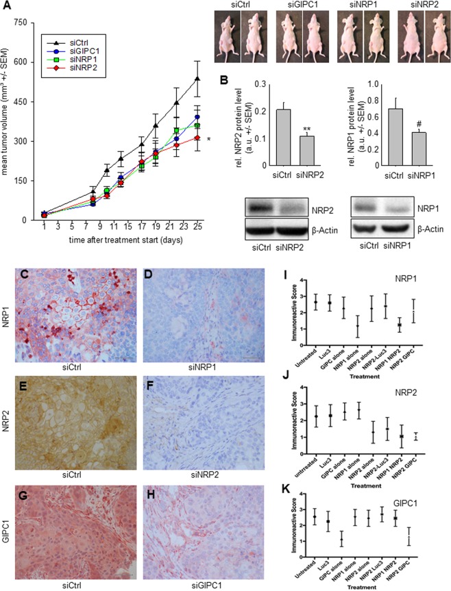
Figure 6.
Therapy study in s.c. xenograft-bearing mice using nanoparticle-formulated siRNAs. (A) Tumor growth curves of Colo357 xenografts upon treatment with PEI-complexed specific or non-specific siRNAs. Right panel: representative examples of mice upon termination of the experiment. (B) Quantitation of NRP2 (left) or NRP1 protein levels (right) in tumor xenografts after explantation. Lower panel: representative Western blots of tumor lysates from tumor xenografts treated as indicated. (C–H) Immunohistochemistry of transplanted tumors, stained with antibodies against the silenced proteins. Representative tissue pieces of tumor xenografts from mice treated with specific siRNA (right panels) vs. negative control treatments (left panels) are shown. Tissues are stained for NRP1 (C,D), NRP2 (E,F) and GIPC1 (G,H). Bar: 2 µm. (I–K) Graphs illustrating the quantitation of immunoreactive staining. Means of the immunoreactive score of the specific staining is given for all treatment groups, with tissues stained for NRP1 (I), NRP2 (J) and GIPC1 (K).

