Figure 3.
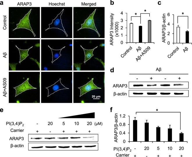
Aβ-mediated SHIP2 activation reduces the expression level of ARAP3. (a) Representative images of ARAP3 (green) in HT22 cells incubated without or with 1 μM Aβ in the absence or presence of 10 μM AS09. Cell nuclei were stained with hoechst33342 (blue). (b) The fluorescence intensity of cytosolic ARAP3 was quantified (means ± SEM; t-test; *p < 0.05; n = 78~100 cells per group). (c,d) The expression levels of ARAP3 in SH-SY5Y cells with or without the treatment of 1 μM Aβ for 24 hr. ARAP3 level was quantified by densitometry analysis and normalized to β-actin (means ± SEM; t-test; *p < 0.05; n = 4). (e) The expression level of ARAP3 in HT22 cells treated by various concentrations of PI(3,4)P2 with or without histone carriers for 24 hr. β-actin was used as a loading control. (f) ARAP3 level was quantified by densitometry analysis and normalized to β-actin (means ± SEM; t-test; *p < 0.05; n = 3).

