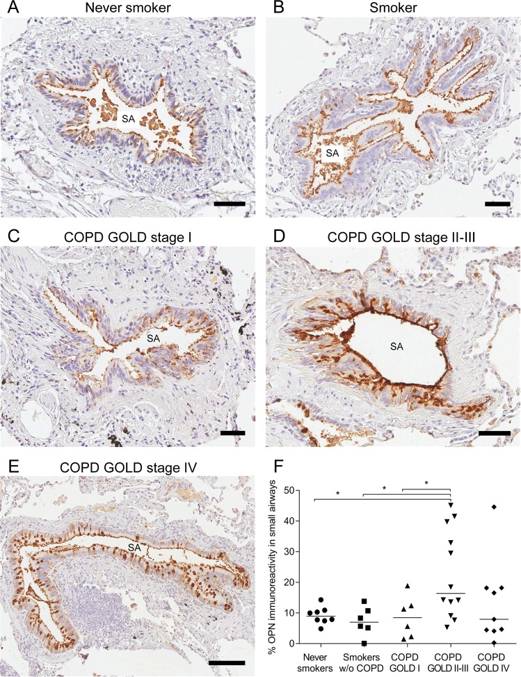
Figure 1.
Localization of osteopontin (OPN) in small airways. Immunoreactivity for OPN was visualized via reaction with 3,3′-diaminobenzidine (DAB), resulting in brownish staining. OPN was observed on the apical surface of the airway mucosa as well as in cells scattered within the epithelial cell layer of small airways in a never-smoker (A), smoker without chronic obstructive pulmonary disease (COPD) (B), and patients with COPD at various stages of the Global Initiative for Chronic Obstructive Lung Disease (GOLD) classification (C–E). Analysis of immunoreactivity within the epithelial cell layer was performed and groups were compared via one-way analysis of variance (ANOVA) with Dunnett’s post-hoc test. (F) *P < 0.05. SA = Small airway. Scale bars: A–D = 50 µm, E = 150 µm.

