Figure 1.
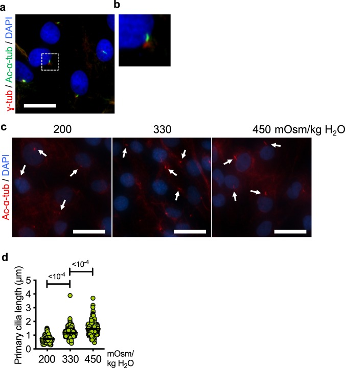
NP cell primary cilia modulate their lengths in response to changes in extracellular osmolarity. (a) Immunofluorescence staining of acetylated α-tubulin (green) and γ-tubulin (red) to mark primary cilia axoneme and basal bodies, respectively, in primary rat NP cells. Scale bar = 30 μm. (b) Zoomed-in image of a primary cilium from the area demarcated by the white square in panel (a). (c,d) Primary cilia of rat NP cells cultured under different osmotic conditions for 24 h were visualized by immunofluorescence staining of acetylated α-tubulin. (c) The lengths of primary cilia increase in response to increased osmolarity (450 mOsm/kg H2O) compared to isoosmotic control (330 mOsm/kg H2O) conditions, whereas they appear shorter under hypoosmotic conditions (200 mOsm/kg H2O). White arrows mark primary cilia. Scale bar = 50 μm. (d) Quantification of primary cilium length was done using ImageJ software. (n = 3 experiments; at least 150 cells/group) Data are represented as scatter plots (mean ± SEM). One-way ANOVA with Dunnett’s multiple comparison test was used to determine statistical significance.

