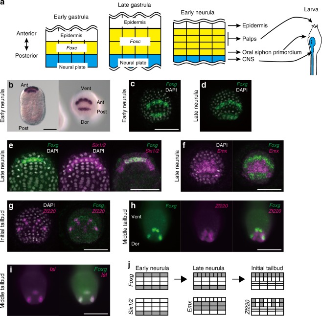
Fig. 1.
Foxg is specifically expressed in two rows of the anterior border of the neural plate. a Schematic illustrations of the anterior border of the neural plate in ascidian embryos. Epidermal cells, neural plate cells, and the intervening cells are represented by white, cyan, and yellow rectangles, respectively. b–d Foxg expression revealed by chromatic and fluorescence in situ hybridization at the early and late neurula stages. Foxg is initially expressed in two separate rows during the neurula stage, and cells in the anterior row divide along the mediolateral axis until the late neurula stage. e–i Double fluorescence in situ hybridization showing expression of e Foxg (green) and Six1/2 (magenta), f Foxg (green) and Emx (magenta), g, h Foxg (green) and Zf220 (magenta), and i Foxg (green) and Isl (magenta) at the late neurula to middle tailbud stages. Photographs are Z-projected image stacks overlaid in pseudocolor. The brightness and contrast of these photographs were adjusted linearly. Nuclei stained by DAPI are shown in gray in some photographs. j Depictions of the expression patterns of Foxg, Six1/2, Emx, and Zf220 in the neural plate border at the neurula stage. b, e Dorsal views in which the anterior is up. c, d, f–i Anterior views in which the ventral side is up. Ant anterior, Post posterior, Dor dorsal, Vent ventral. Scale bars represent 50 μm

