Fig. 3.
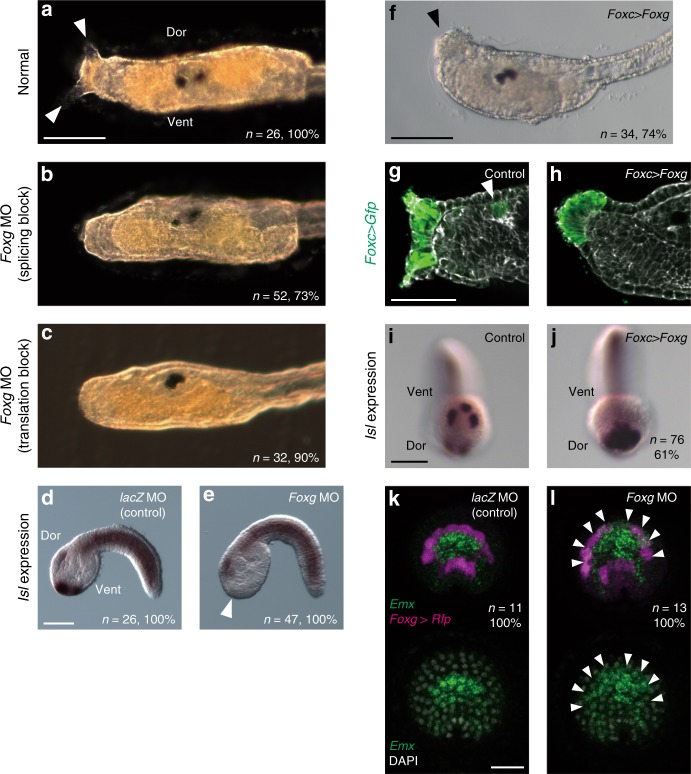
Foxg is essential for palp formation. a–c Morphology of the larval trunk developed from a control unperturbed eggs, b eggs injected with a MO designed to block splicing of Foxg RNA, and c eggs injected with a MO designed to block translation of Foxg mRNA. While two of three palp protrusions are visible in normal embryos shown in (a) (arrowheads), no palp protrusions are visible in the morphants shown in (b) and (c). d, e Isl expression in embryos injected with d the control lacZ mRNA and e the Foxg MO. The palp-forming region lost Isl expression in the Foxg morphant shown in (e) (arrowhead). Lateral views are shown. f A larva in which Foxg was over/misexpressed using the upstream region of Foxc developed a single large palp (arrowhead). g, h The ANB cells were marked with GFP by introducing the Foxc > Foxg construct and counterstained with phalloidin (gray). The anterior trunk regions of g a control larva and h a larva in which Foxg was over/misexpressed are shown. g In normal embryos, ANB cells contribute to the palp region and the oral siphon primordium (arrowhead). h In embryos with Foxg over/misexpression, all Gfp-positive cells were found in the single large palp. i, j Isl expression in (i) a control embryo and (j) an embryo with Foxg over/misexpression. Isl is expressed in the entire palp-forming region in (j). k, l Fluorescence in situ hybridization for Emx and Rfp. Embryos were injected with k the lacZ MO or l the Foxg MO. Foxg > Rfp was coinjected to mark the most anterior and posterior rows of the ANB and detected by in situ hybridization. In (l), ectopic expression of Emx was detected in the anterior row. The number of embryos examined and the proportion of embryos that each panel represents are shown within the panels. The brightness and contrast of fluorescence images were adjusted linearly. Scale bars represent 50 μm

