Fig. 8.
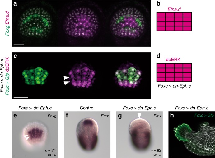
Efna.d is responsible for restricting Foxg expression in the ANB. a Double in situ hybridization of Efna.d (magenta) and Foxg (green). b Depiction for Efna.d expression in the ANB. c Immunostaining of dpERK (magenta) in an embryo in which ANB cells were marked with Foxc > Gfp. The middle rows show ectopic signals for dpERK (arrowheads). d Depiction of ANB cells stained with the anti-dpERK antibody in neurula embryos introduced with Foxc > dn-Eph.c. e–g In situ hybridization of e Foxg and f, g Emx in e, g neurula embryos introduced with Foxc > dn-Eph.c and f an unperturbed neurula embryo. In (e), the middle two rows of cells expressed Foxg ectopically (compared with Fig. 1b). In (f) and (g), while Emx expression in the tail was not changed, it was lost in the ANB of a Foxg morphant (arrowhead). The number of embryos examined and the proportion of embryos that each panel represents are shown within the panels. a, c, e Anterior views in which the ventral side is up. f, g Dorsal views in which the anterior is up. h Lateral view showing the morphology of a larva introduced with Foxc > dn-Eph.c. ANB cells were marked with GFP by cointroduction of the Foxc > Gfp construct and counterstained with phalloidin (gray)

