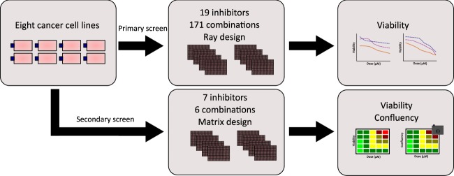
Fig. 1.
Schematic representation of study design. Eight human cancer cell lines from different tissue origins were used in this study. Cells were incubated overnight prior to drug addition. In the primary screen, cells were screened against 19 small-molecule inhibitors in single and double application, with a total of 171 combinations. After 48 hours of drug exposure the assay was terminated, and cell viability was measured using CellTiter-Glo 2.0 (Promega). In the secondary screen, cells were screened against 7 single small-molecule inhibitors and 6 combinations for a duration of 48 hours. Drug effect was measured using automated brightfield imaging of confluency and CellTiter-Glo 2.0 (Promega).

