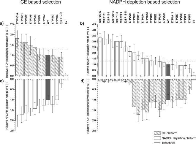
Figure 4.
Rescreening of variants from MP-CE based selection, (a,c), and NADPH depletion based selection (b,d). Identified P450 BM3 variants from both screening methods were measured in triplicates and compared to the P450 BM3 WT (grey column) and EV (negative control). Variants which showed an increase of factor ≥1.3 (2.5-fold standard deviation, dashed line) in 4-hydroxy-isophorone formation (dashed columns) within the CE based selection (a) or NADPH oxidation rate (white columns) within the NADPH depletion based selection (b) were selected for further characterization. For a full picture, variants from the CE based selection (c) and NADPH depletion based selection (d) were screened with the opposite platform, however, results not considered for selection. (measurements: n = 4, mean ± s.e.; in biologically independent experiments) Substitutions of variants see Supplementary Table S2.

