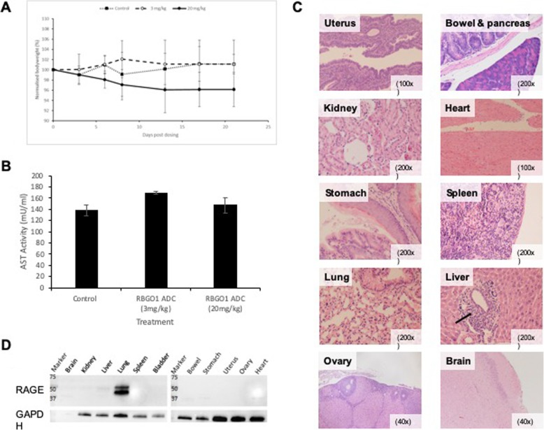
Fig. 6.
RBGO1 ADC is not toxic in a murine in vivo model. PBS (Control) or RBGO1 ADC was administered (intravenously) to female, athymic mice aged 5–7 weeks and weighing approximately 28-35 g, at a dose of either 3 mg/kg or 20 mg/kg. Bodyweight a was measured at days 3, 6, 8, 13, 17 and 21 and mice were sacrificed at either 24 h or 3 wks following dosing, after which full blood counts and an aspartate aminotransferase (AST) ELISA were performed (b). Organs were harvested immediately following sacrifice, and formalin fixed and paraffin embedded before sectioning and staining with hematoxylin and eosin (c). Western blot analysis of mouse tissue was performed using the RBGO1 antibody (d). Representative images were acquired on a Zeiss Axio Imager 2 microscope and analyzed using the ZEN 2012 image analysis software and magnifications are shown on each image. Low level inflammatory cell infiltration is indicated in the ‘Liver’ image (⟶). Data displayed in histograms are means of three animals. Data were analyzed by ANOVA and Dunnett’s multiple comparison test. ADC treatments differ from control, **p < 0.01

