
Fig. 1.
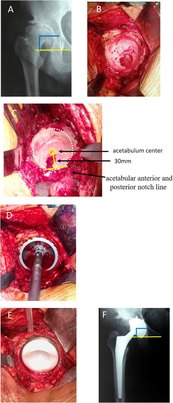
Radiographs of a 39-year-old female patient with end-stage osteoarthritis secondary to Crowe type I DDH. a Preoperative X-ray. b Intraoperative image showed shallow cup-shaped acetabulum and crack-shaped Harris fossa. c Locating the acetabulum center. d Reaming the acetabulum and installing the test cup. e Installing the acetabular prosthesis. f Postoperative X-ray and rotation center restoration