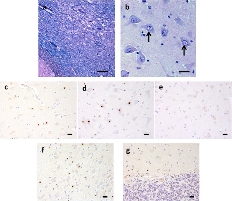
Fig. 1.

Neuropathology of FXTAS. Luxol fast blue-H&E stain shows spongiosis of the cerebellar white matter (a). Abundant eosinophilic intranuclear inclusions (arrows) were found in neurons of the cerebral cortex, especially in hippocampal pyramidal cells (b). Intranuclear inclusions in the CA4 region of the hippocampus were immunorective for p62 (c), NTF1 (d) and CTF1 (e). Neurons and glia in pontine nuclei (f NTF1), as well as Bergmann glia and rare Purkinje neurons of the cerebellum also contained intranuclear inclusions (g NTF1). [Scale bar = 100 μm in a; 20 μm in b, c, d, e, f and g]
