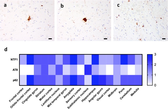
Fig. 2.
PSP-like changes were detected by AT8 immunohistochemistry as tufted astrocytes (a), globose tangles (b) and coiled bodies (c). (d) Heatmap of relative abundance of NTF1-positive pathology (upper row), AT-8 positive pathology (middle row) and p62-positive pathology (lower row). 0 = no pathology, 1 = mild pathology, 2 = moderate pathology and 3 = severe pathology). Scale bar: 20 μm

