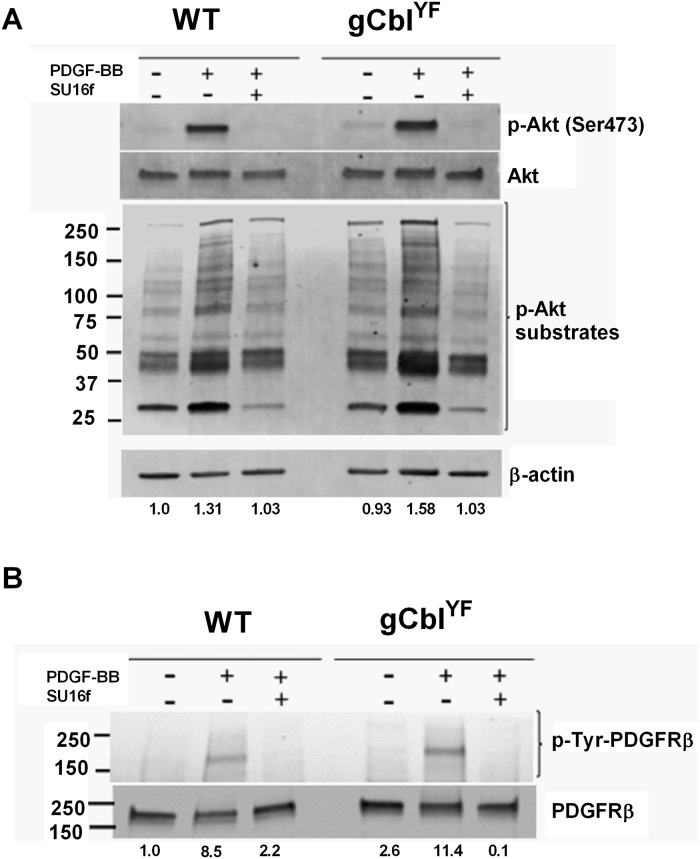
Fig 3. PI3K upregulation sensitizes periosteal cells to the effects of PDGF.
Periosteal cells isolated from WT or gCblYF mice were serum starved for 1–2 hours and then treated with PDGF-BB (10ng/mL) or SU16f (5μM), a PDGFR inhibitor, for 30 minutes. (A) Total cell lysate (40 μg) was electrophoresed on 10% SDS-PAGE. Western blots were probed with anti phospho-Akt antibodies and anti-Akt antibodies. Another set of blots were probed with anti-Akt substrate antibody (top), and then stripped and reprobed with anti-β-actin antibodies (bottom) to verify equal loading of protein. The ratio of phosphorylated and total protein is indicated at the bottom of each lane. (B) The blot was probed using anti-phospho-tyrosine antibody (top), to determine phosphorylation of the PDGFR, and then stripped and reprobed with anti-PDGFRβ (bottom) to verify equal loading of protein. The ratio of phosphorylated and total protein is indicated. A representative of two experiments is shown.

