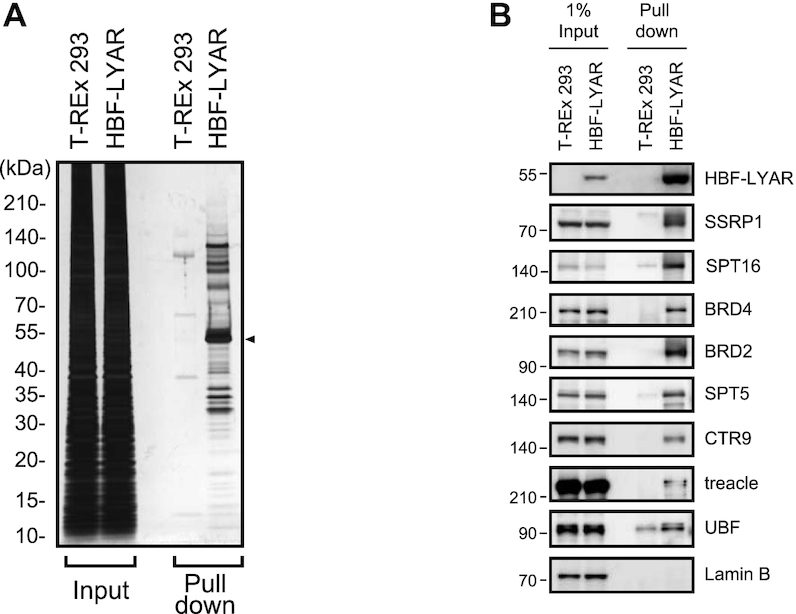
Figure 2.

Pulldown of LYAR and identification of LYAR-associated proteins (A) Silver staining of HBF-LYAR-associated proteins. HBF-LYAR-associated complexes were isolated via sequential two-step pulldown (Ni-NTA pulldown, RNase A treatment, and pulldown of FLAG-tagged HBF-LYAR) from nuclear extract of HBF-LYAR-TO cells or T-REx 293 cells (control) treated with Dox for 24 h. Proteins were subjected to SDS-PAGE and visualized with silver staining. The arrowhead represents HBF-LYAR, as the bait protein. Molecular mass markers (kDa) are indicated to the left. Input: nuclear extract (10 μg). (B) Immunoblotting of HBF-LYAR-associated proteins using antibodies indicated to the right of each panel. 1% Input: 1% of the nuclear extract used for pull down of HBF-LYAR complexes. HBF-LYAR was detected by Stabilized Streptavidin-HRP Conjugate.
