Figure 3.
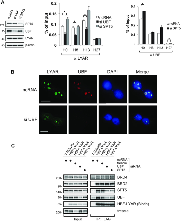
LYAR recruitment to rDNA loci depends on UBF (A) ChIP analysis of LYAR (left) or UBF binding (right) to rDNA loci in 293T cells upon knockdown of UBF or SPT5. 293T cells were treated with an siRNA specific for UBF or SPT5 (ncRNA as a control) for 72 h, and whole-cell extract was subjected to immunoblotting using the antibodies indicated to the right of the panels. β-actin was used as the loading control. Molecular mass markers (kDa) are indicated to the left. The graphs show the amount of ChIPed DNA (% of input) with respect to particular rDNA loci. Data represent the mean ± SEM of three independent experiments. *P < 0.05 (unpaired t-test). (B) Immunocytostaining of LYAR and UBF upon UBF knockdown. 293T cells were treated with ncRNA or an siRNA specific for UBF for 72 h and subjected to immunocytostaining with anti-LYAR (rabbit) and anti-UBF (mouse). FITC-conjugated anti-rabbit IgG and Cy3-conjugated anti-mouse IgG were used as the secondary antibodies. DAPI staining indicates the nucleus. Bar: 10 μm. (C) Immunoblotting for HBF-LYAR-associated proteins upon siRNA-mediated knockdown of treacle, UBF or SPT5 in HBF-LYAR-TO cells for 72 h. A two-step pulldown was carried out with His6- or FLAG-tag of HBF-LYAR. The antibodies used for immunoblotting are indicated to the right.

