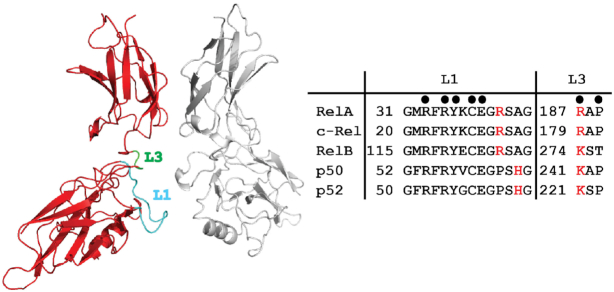
Figure 4.
NF-κB residues that participate in sequence-specific DNA contacts. Left, ribbon diagram representation of RelA homodimer (PDB ID: 2RAM) highlighting loops L1 (cyan) and L3 (green). These two loops contribute all amino acid side chains that contact DNA bases. Right, the amino acid sequences and numbering for loops L1 and L3 are shown for all five murine NF-κB subunits. Filled circles above mark positions of identically conserved DNA base-contacting residues. Red letters correspond to nonidentical residues that either participate in DNA base-contacts in all cases (denoted by circle) or in specific circumstances, as described in the text.

