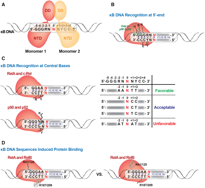
Figure 5.
Variable modes of NF-κB:DNA recognition. (A) NF-κB binds to κB DNA as a dimer and each monomer recognizes one half site. (B) Standard recognition of κB DNA at the 5′-end by NF-κB. The conserved NF-κB subunit loop L1 residues involved in direct contacts with DNA bases are indicated. Arg35 (murine RelA numbering) contacts the G base at the -4 position and Arg33 contacts the G at -3. His64 or His62 are specific only to p50 and p52 subunits, respectively. (C) NF-κB recognition of κB DNA at the central bases. Left, different modes of DNA binding (for RelA and c-Rel vs. p50 and p52) are shown. Right, table summarizing how the presence of specific nucleotides at certain central positions affect NF-κB:DNA binding. (D) κB DNA sequence induces NF-κB binding with distinct modes. R41/R125 of RelA/RelB binds the G:C bp at –5 position of a 5 bp half-site. R187 of RelA cannot bind the –2 position of the same half-site.

