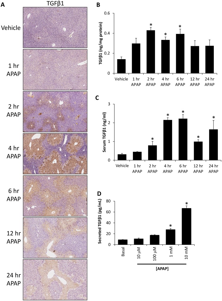
Figure 2.
Hepatic TGFβ1 signaling was increased in APAP-treated mice. A, Images of immunohistochemistry for TGFβ1 in liver sections from vehicle and time course APAP-treated mice. B, TGFβ1 concentration in liver homogenates from vehicle and APAP-treated time course mice as measured with ELISA. C, Serum TGFβ1 levels as measured by ELISA in samples from vehicle and time course APAP-treated mice. D, TGFβ1 concentration measured by ELISA in conditioned media from primary hepatocytes treated with the indicated doses of APAP. *p < .05 compared with vehicle-treated mice.

