Figure 4.
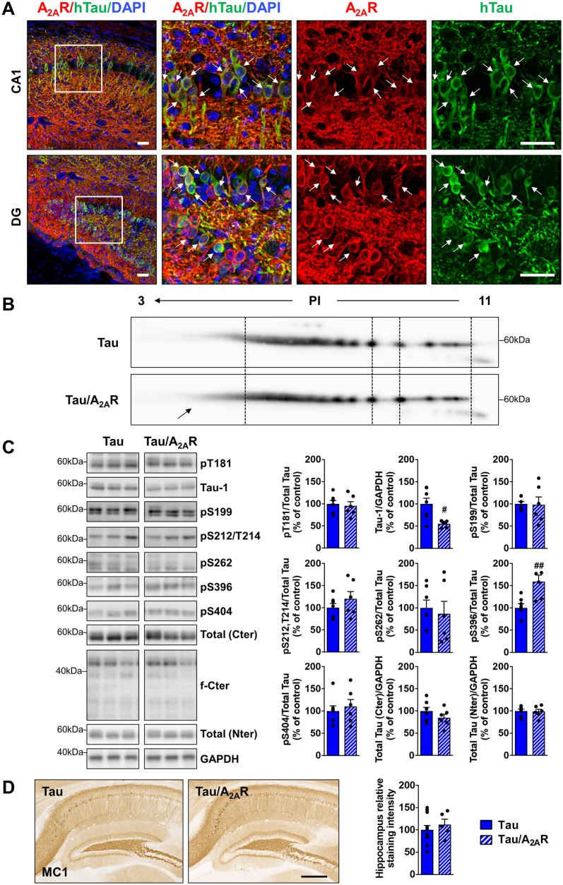
Impact of neuronal A2AR overexpression on hippocampal tau pathology. Human tau expression, phosphorylation and aggregation in the hippocampus of triple transgenic mice (tau/A2AR) versus tau transgenic controls were evaluated by immunohistochemistry, bidimensional electrophoresis (2D) and western blots. (A) Co-immunostainings with A2AR (red) and human tau (TauE1E2 antibody, human total tau, green) in the CA1 and dentate gyrus (DG) regions of triple tau/A2AR transgenic mice. Neurons expressing human tau transgene (arrows) were found to overexpress A2AR. DAPI (blue) represents cell nuclei. Scale bar = 50 µm. (B) 2D profile of total human tau (Cter antibody) in triple tau/A2AR mice and littermate tau controls, shows an increase of tau isovariants in the acidic range of PI (arrow). (C) Quantification of tau phosphorylation at T181, S199, S212/T214 (AT100), S262, S396 and S404 epitopes, as well as dephosphorylated tau (tau-1) in triple tau/A2AR animals and littermates tau controls. Analysis revealed tau hyperphosphorylation in tau/A2AR mice signed by increased pS396 and reduced tau-1 (dephosphorylated tau). #P < 0.05, ##P < 0.01 versus tau mice using Student’s t-test. n = 6–7 per group. (D) Conformational tau immunostaining using MC1 antibody in triple tau/A2AR animals and littermates tau controls revealed no difference between groups. n = 5–11 per group. Scale bar = 500 µm. Results are expressed as mean ± SEM.

