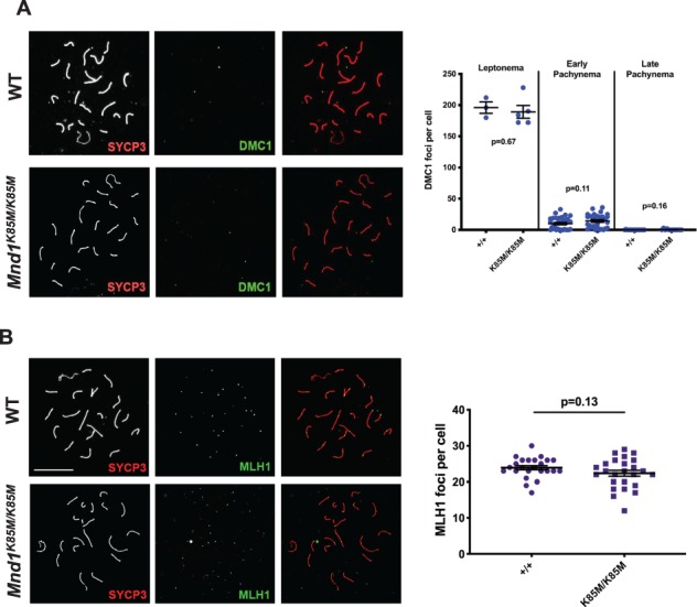
Figure 3.

Cytological data indicating that Mnd1K85M/K85M is not defective in meiotic recombination. (A) Late pachytene spermatocyte chromosome surface spreads immunolabeled with synaptonemal complex axial element protein (SYCP3) and DMC1 (merged in rightmost subpanels). To the right of each panel are plots of DMC1 focus counts at indicated substages. N = 2 per genotype. P values are from Student’s t-test. (B) Spreads immunolabeled with MLH1 (a chiasmata marker protein) and SYCP3 (merged in rightmost subpanels). Size bar in WT panel of B = 20 μm, and applies to all panels. To the right of each panel are plots of the MLH1 focus counts at indicated substages. N = 3 per genotype. DMC1: dosage suppressor of mck1 homolog.
