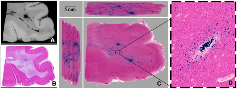
Figure 5.
Pathology underlying TMBs: injury to the vasculature. (A) High-resolution 7 T MRI of tissue block containing region of interest co-localized to TMBs observed in vivo and ex vivo whole-brain MRI. (B) Standard 2D histopathology section (20-μm thick) from the imaged tissue block (A) stained with Perls Prussian Blue reveals iron in macrophages surrounding a large vessel. (C) Serially cut tissue sections digitized as a 3D volume, shown as side views in the adjacent panels. A 3D minimum intensity projection (MIP) image of the Perls stained histology reconstructed to match high-resolution 7 T MRI (A). (C) 3D reconstruction of interleaved tissue sections stained for iron deposits demonstrates iron laden macrophages that are connected and branching over centimetres of tissue. (D) Enlarged section of (C) showing iron-laden macrophages that co-localized with a vessel. These interleaved serial sections of the TMB in tri-planar view reveal a territory of injured vasculature in areas visible on MRI as punctate or linear hypointensities. Injury to the large vessel is indicated by confluent regions of iron-laden macrophages that create sufficient signal for detection on MRI. Injury to some of the other smaller vessels is only detected on post-mortem histology in a 3D reconstruction. The tri-planar view and high resolution identifies connectivity of the injured vessels ranging from large to small diameters.

