
Fig. 1. LI-rTMS induces transcommissural climbing fiber reinnervation in pedunculotomized adult mice.
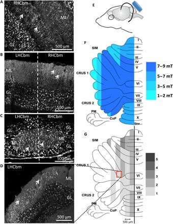
(A) Climbing fibers (white arrows) in the molecular layer (ML) of the intact right hemisphere (CRUS 1 and CRUS 2 coronal sections) from a sham-treated mouse (n = 5). (B) After sham treatment, in coronal sections, VGLUT2-positive climbing fibers (white arrows) in the molecular layer of the right hemicerebellum stop at the midline (thick vertical dashed line), consistent with lack of reinnervation (18). (C) In comparison after LI-rTMS (n = 9), VGLUT2-positive climbing fibers (white arrows) fill the molecular layer on both sides of the midline (thick vertical dashed line) in this coronal section, consistent with reinnervation. (D) Further laterally, VGLUT2-positive climbing fibers (white arrows) in the molecular layer of the lesioned hemisphere following LI-rTMS. Anatomical differences from (A) and (B) reflect the noncoronal orientation of the lobule (simplex), and therefore, climbing fiber arbors are angled to the coronal plane of the section. (E) Diagram showing the coil (blue) in relation to the mouse head. (F) Unfolded cerebellum showing magnetic field intensity delivered by LI-rTMS, as measured by a Hall device in air at corresponding X, Y, and Z distances from the center of the coil. (G) Average density, in 0.5-mm parasagittal zones, of LI-rTMS–induced climbing fiber reinnervation (n = 9). This parasagittal organization of different reinnervation densities is consistent with that previously demonstrated in BDNF-induced climbing fiber reinnervation, which demonstrates parasagittal topography and provides recovery of motor and navigation behaviors (18). Grayscale grading: 1 = few strands; 2 = one-quarter lobule; 3 = half lobule; 4 = three-quarter lobule; and 5 = completely climbing fiber–filled (18). GL, granular layer; LHCbm, left hemicerebellum; RHCbm, right hemicerebellum; SIM, lobulus simplex; PM, paramedian lobule; CoP, copula pyramidis; I to X, lobules 1 to 10 of the vermis. Photo credit: A. D. Tang, University of Western Australia, Experimental and Regenerative Neuroscience Laboratory.