. 2019 Sep 24;8:e48958. doi: 10.7554/eLife.48958

Figure 6. Stem cell activity depends on cycling cells but is not confined to cells from the neck.

(a-b) DAPI-stained worms after rescue with cell transplantations from whole-worm donors (a) or sourced from depicted donors (b). (c) Quantification of rescue phenotypes from pooled experiments. Number of animals listed above bars. (d) Model for head-dependent neck maintenance and proglottid formation. (e) Models of head-dependent or -independent stem cell niches.

Figure 6.

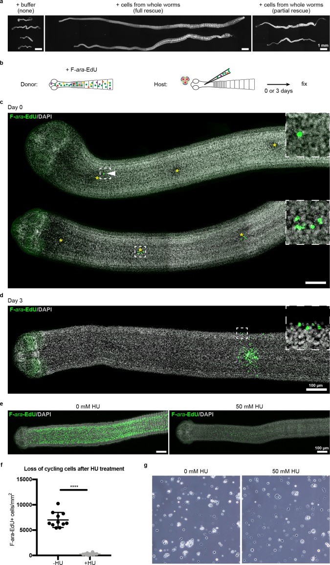
Figure 6—figure supplement 1. Stem cell activity depends on cycling cells.

Figure 6—figure supplement 1.

(a) DAPI-stained worms showing phenotypes observed after attempted rescue of irradiation-induced lethality. No rescue results in degenerated worms with no proglottids, full rescue results in normal worms with multiple proglottids, and partial rescue refers to worms with visible proglottids but with defects such as contracted necks. (b) Schematic for rescue experiment using donors with labeled cycling cells. (c–d) Maximum-intensity projections of tile-stitched confocal sections 0 or 3 days post-transplantation according to (b). Injections sites marked with asterisks. White arrowhead points to a single transplanted cell. After 3 days, large colonies of F-ara-EdU+ (green) cells could be detected with some labeled cells incorporated into terminally differentiated tissues at the animal edge (inset). (e) Maximum-intensity projections of tile-stitched confocal sections after 1 hr F-ara-EdU uptake (green) from control worms or worms cultured with hydroxyurea (HU) for 6 days. (f) Quantification of cycling cells from (e). Error bars = SD, N = 3, n = 11 and 8, Student’s t-test. (g) Cell morphology with or without HU treatment prior to transplantation.

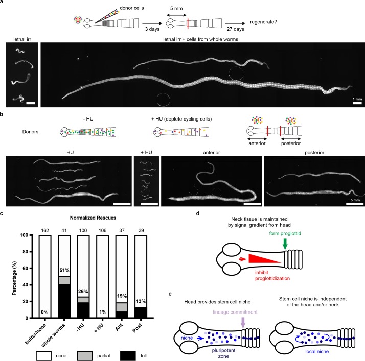
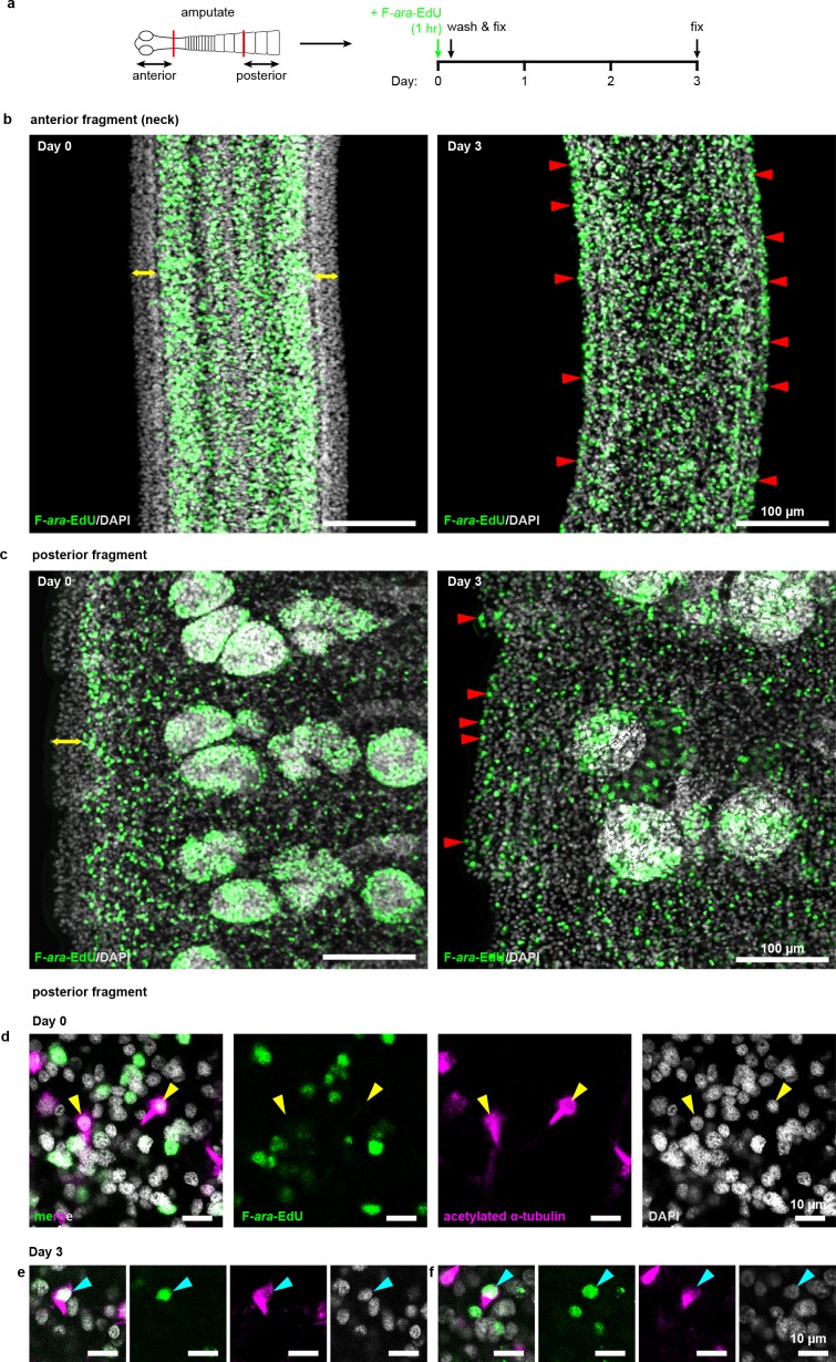
Figure 6—figure supplement 2. Cycling cells give rise to multiple lineages in both anterior and posterior fragments.

Figure 6—figure supplement 2.

(a) Schematic of F-ara-EdU pulse-chase protocol to label cycling cells from 2 mm amputated fragments and detect their progeny after 3 days. (b–c) Confocal sections of both anterior and posterior fragments on day 0 showing the absence of F-ara-EdU (green) at the animal edge (yellow double-headed arrows) where differentiated muscle and tegument are located. After 3 days, F-ara-EdU has chased into the edge-most nuclei (examples marked by red arrowheads). (d-f) Confocal sections from posterior fragments after F-ara-EdU pulse-chase in combination with acetylated α-tubulin antibody staining to label flame cells (magenta). Yellow arrowheads point to F-ara-EdU- flame cell nuclei at day 0 (d) which are exclusively post-mitotic. Cyan arrowheads point to two examples of flame cell nuclei that are F-ara-EdU+ after 3 days chase (e–f).