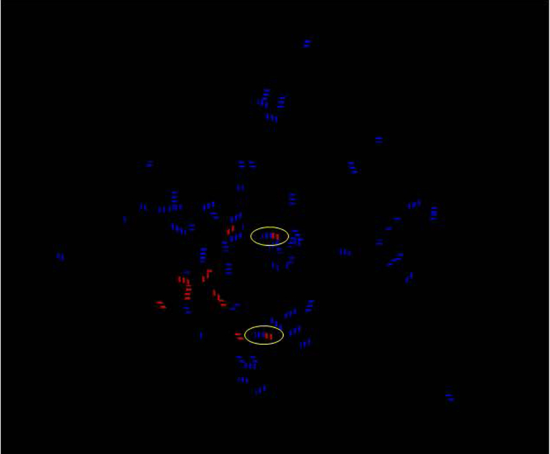
FIGURE 6.
Hydrogen bonding analysis performed in VMD using chain coloration demonstrate at least two potential hydrogen bonds between the BNP target and each of the aptamers (circled blue-red hybrid dashed lines) and potential stabilizing intra-aptamer hydrogen bonds shown as blue dashed lines. Purely red dashed lines represent hydrogen bonds within BNP itself. Of course, these analyses were performed in vacuo without consideration for the abundant aqueous solvent, although water could interfere with any of the possible hydrogen bonds or exclusion of water during aptamer-ligand could be a thermodynamic (positive entropy change or + ΔS) driver for BNP-aptamer binding.11

