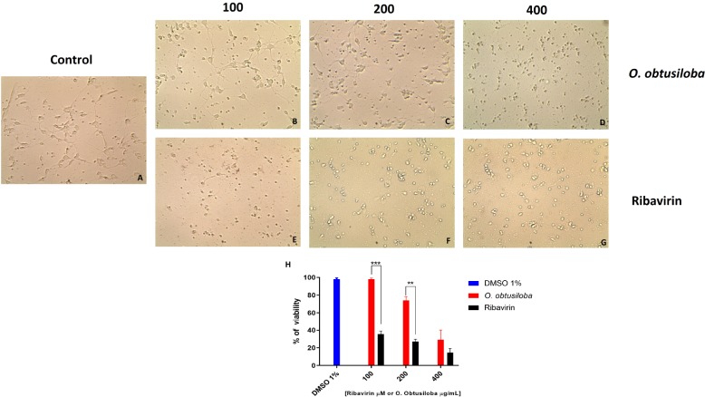
FIGURE 2.
Cytotoxicity of O. obtusiloba extract and ribavirin in chick retina neurons in 4-day-old purified cultures. (A) Control culture. (B,C,D) Cultures incubated with O. obtusiloba at concentrations of (B) 100 μg/mL, (C) 200 μg/mL and (D) 400 μg/mL. (E,F,G) Cultures incubated with ribavirin at concentrations of (E) 100 μM, (F) 200 μM and (G) 400 μM. (H) Graphical representation showing the percentage of viability of treated cells with O. obtusiloba (blue bar) and percentage of viability of treated cells with ribavirin (dark bar). The results were evaluated by MTT assay and showed that the exposure to the O. obtusiloba extract presented low cytotoxicity maintaining high viability in concentrations up to 200 μg/mL. Error bars indicate the standard deviation and experiments were performed in triplicate. ∗∗p < 0.01; ∗∗∗p < 0.001 in Tukey test.

