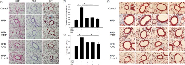
Figure 3.
Histopathological assessment using haematoxylin and eosin (H&E), periodic acid-Schiff (PAS), and Mason’s trichrome (MT) staining (×200) (A), quantitative fibrosis area assessed using MT staining (B), and hydroxyproline level (C) among groups. Fibrosis was more predominantly observed in peribronchial and perivascular area compared that in lung parenchymal lesion in MT staining (×400) (D). One-way ANOVA followed by a post-hoc Bonferroni test was used in Fig. 4B,C. HFD, high-fat diet; EMP, empagliflozin; DUL, dulaglutide; combi, co-treatment with EMP and DUL.

